Data collection
Study design
Twenty healthy adults (mean age 36 years, age range 29-46 y.o.(SD = 4.7), 4 men and 16 women) were scan at the Centre Hospitalier Universitaire of Sherbrooke (CHUS) using a clinical 3T MRI scanner (Ingenia, Philips Healthcare, Best, Netherlands) with a 32-channel head coil. Each MRI session was repeated 5 times over 5 months and a 4-week interval (+/- 1 week). For each participant, images were acquired at approximately the same time of day to avoid potential diurnal effects (i.e., a morning participant had all sessions in the morning, with a tolerated 2–3-hour variation).
Study design - 20 healthy subjects

All MRI data acquisitions were aligned on the anterior commissure-posterior commissure plan (AC-PC) and each MRI session include :
Anatomical 3D T1-weighted (3DT1)
Multi-shell diffusion-weighted images (DWI)
Reverse phase encoding B0 (revb0)
inhomogeneous magnetization transfer (ihMT)
MRI acquisition parameters
Parameters / Sequences |
T1 |
DWI |
Reverse B0 |
ihMT |
T1 ihMT |
|---|---|---|---|---|---|
Phase-encoding direcion* |
RL |
PA |
AP |
RL |
RL |
Technique - Fast imaging method |
FFE - TFE |
SE - EPI |
SE - EPI |
FFE - EPI |
FFE - EPI |
Total scan duration |
4 min 20s |
9 min 20s |
14s |
6 min 04 s |
13 s |
Repetition time (TR, ms) |
7.9 |
4800 |
4800 |
112 |
20 |
Echo Time (TE, ms) |
3.5 |
92 |
92 |
3.6 (Δ = 6) |
3.6 (Δ = 6) |
Inversion Time (TI, ms) |
950 |
||||
Flip Angle (degree) |
8 |
90 |
90 |
15 |
30 |
Field of View (FOV, mm) |
224 x 224 |
224 x 224 |
224 x 224 |
224 x 224 |
224 x 224 |
Slices (n) |
150 |
66 |
66 |
65 |
65 |
Voxel size (mm) |
1 x 1 x 1 |
2 x 2 x 2 |
2 x 2 x 2 |
2 x 2 x 2 |
2 x 2 x 2 |
n b0, b-value (n directions) |
7, 300 (8), 1000 (32), 2000 (60) |
||||
MT stauration pulse |
10 Hann pulses of. 0.9 ms duration with 1.5 ms interval |
||||
Frequency offset of +/- |
7000 Hz |
||||
n Echoes - Echo spacing |
3 - 6 ms |
*The directions are specified in the standard way i.e. in coordinates of the patient (LPH).
An example of bvec and bval file can be downloaded here:
Data conversion: DICOM to BIDS
To convert data we use BIDS standard. An example of the data structure for one subject is shown below:
data-subject
├── dataset_description.json
├── participants.json
├── participants.tsv
├── sub-001_ses-01
├── sub-001_ses-02
├── sub-001_ses-03
├── sub-001_ses-04
├── sub-001_ses-04
├── sub-002_ses-01
├── ...
├── sub-003_ses-01
    │
    ├── anat
    │   ├── sub-003-01_T1w.json
    │   ├── sub-003-01_T1w.nii.gz
    │   ├── sub-003-01_acq-pos_ihmt.json
    │   ├── sub-003-01_acq-pos_ihmt.nii.gz
    │   ├── sub-003-01_acq-neg_ihmt.json
    │   ├── sub-003-01_acq-neg_ihmt.nii.gz
    │   ├── sub-003-01_acq-altnp_ihmt.json
    │   ├── sub-003-01_acq-altnp_ihmt.nii.gz
    │   ├── sub-003-01_acq-altpn_ihmt.json
    │   ├── sub-003-01_acq-altpn_ihmt.nii.gz
    │   ├── sub-003-01_acq-T1w_ihmt.json
    │   └── sub-003-01_acq-T1w_ihmt.nii.gz
    │
    └── dwi
        ├── sub-003-01_dwi.bval
        ├── sub-003-01_dwi.bvec
        ├── sub-003-01_dwi.json
        ├── sub-003-01_dwi.nii.gz
        ├── sub-003-01_b0.json
        ├── sub-003-01_b0.nii.gz
        ├── sub-003-01_rev-b0.json
        └── sub-003-01_rev-b0.nii.gz
To convert our DICOM data folder to the compatible BIDS structure, we used dcm2bids.
dcm2bids -d DICOM_folder -p id_subject -c config.txt -o sub-id
Quality Control raw data
Quality control of raw data was performed using DMRIQC flow DMRIQC flow.
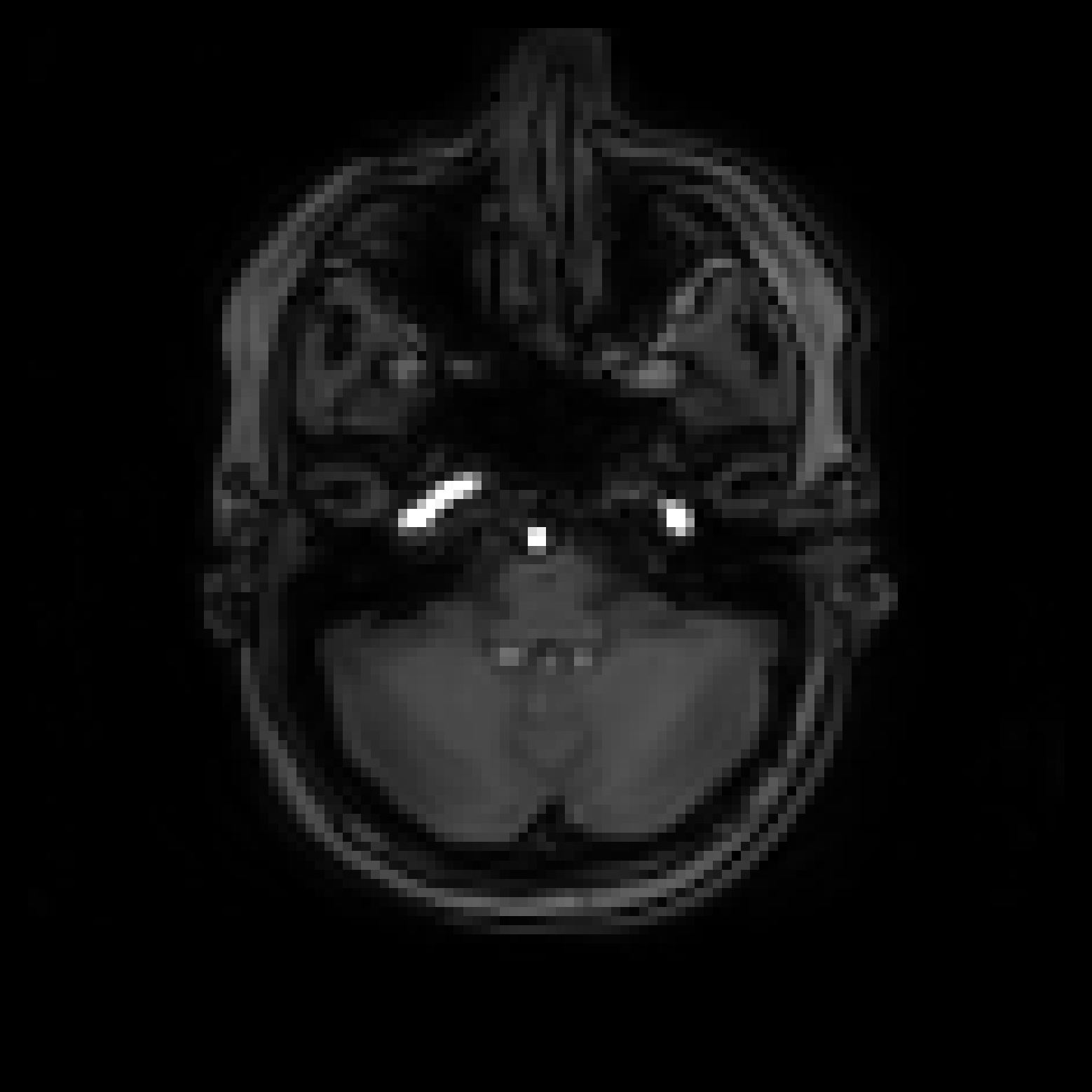
Example of datasets for one subject
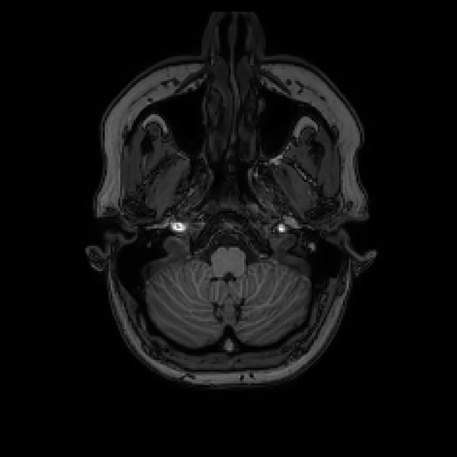
Anatonimal image
3D-T1w |
|---|
|
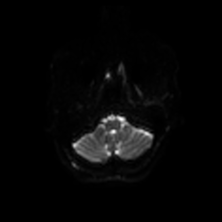
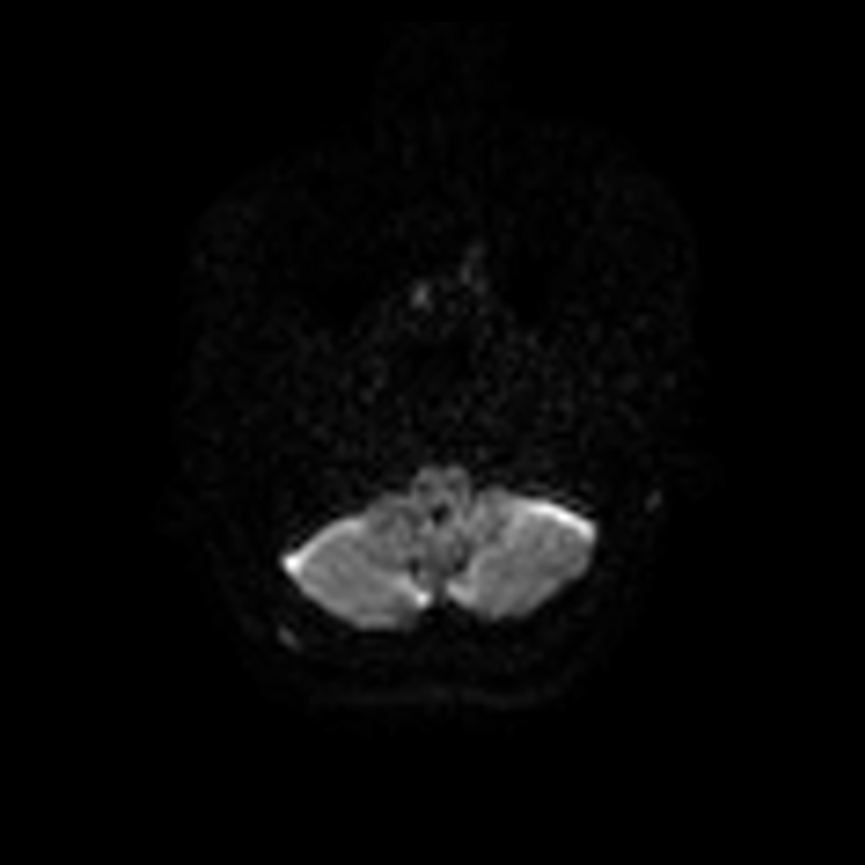
Diffusion images
DWI - b0 |
DWI - b value = 300 |
DWI - b value = 1000 |
DWI - b value = 2000 |
DWI - Reverse B0 |
|---|---|---|---|---|
|
|
|
|

|
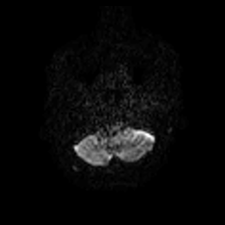
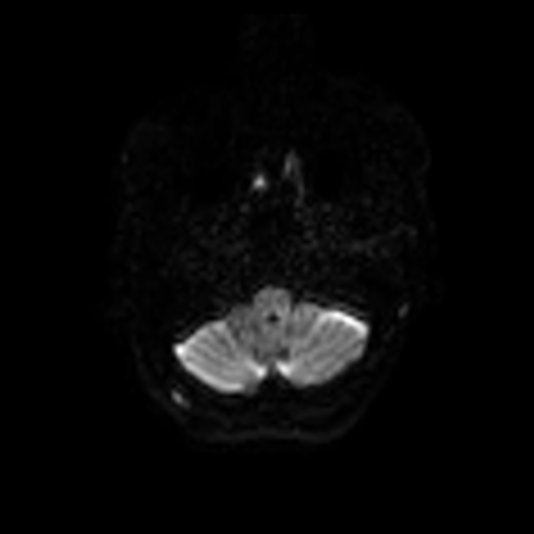
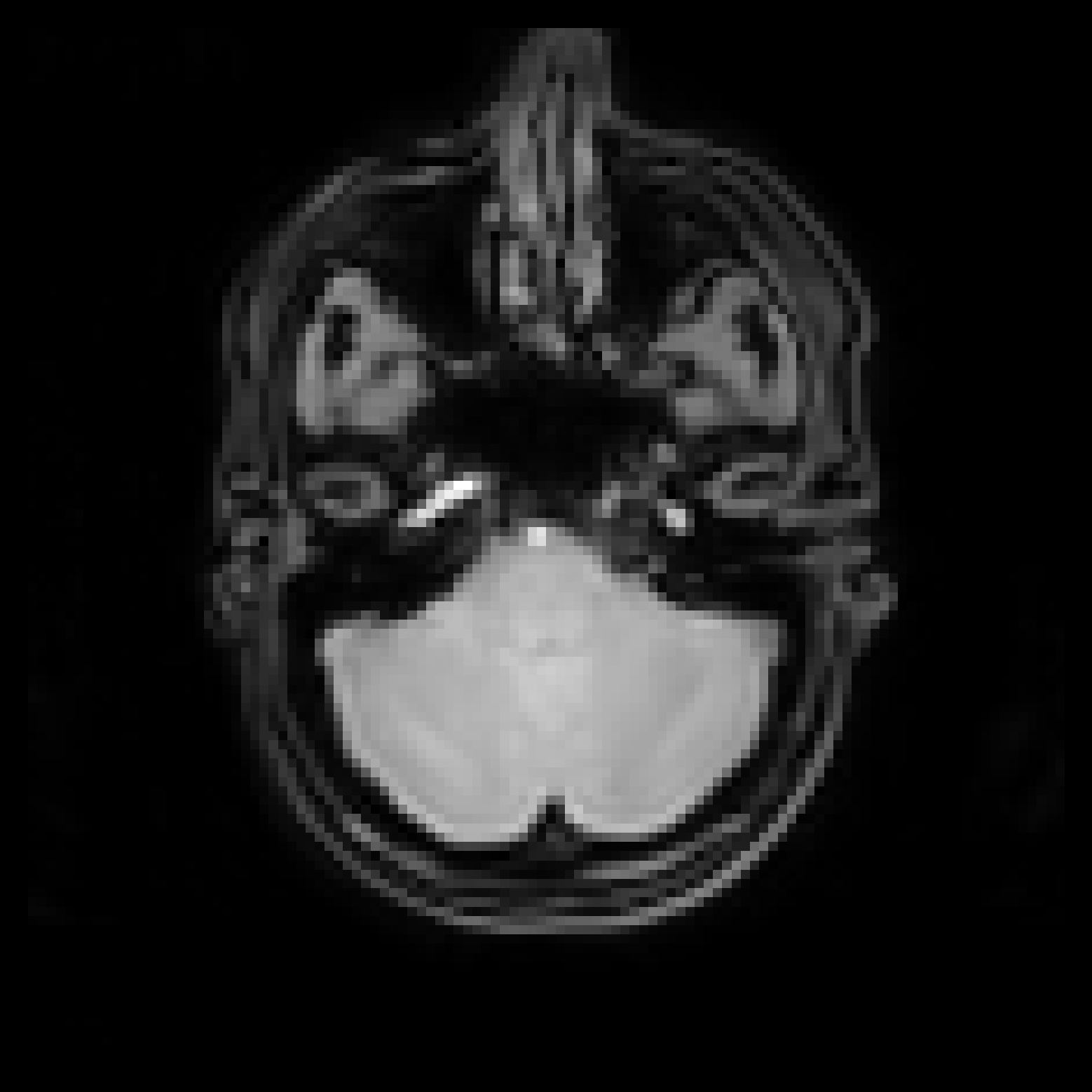
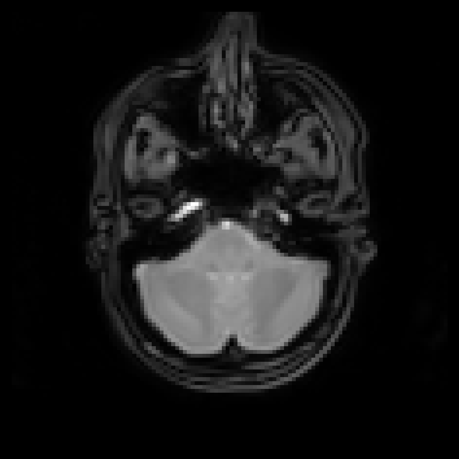
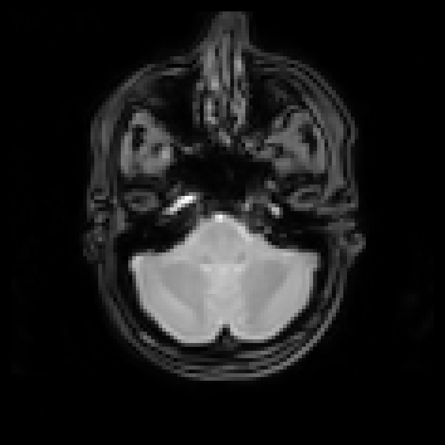
ihMT images
MT-Off |
Positive (pos) |
Negative (neg) |
Alternative pos-neg |
Alternative neg-pos |
T1w ihMT |
|---|---|---|---|---|---|
|
|
|

|

|
|